These programs employ a non-linear least-squares analysis to iteratively determine the best fit for raw data. This is done by changing the positions, heights, and SDs of the Gaussian peak representing sequential daughter generations. Precursor Frequency and Proliferative Indice are two of the most common measures of proliferation. ModFit defines PI as a measure that measures the increase in cell numbers over time. It is similar to the the’stimulation indicator’ for a thymidine-uptake assay.
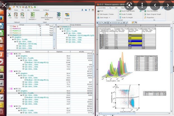
Tree Star Flowjo X10
Password 123
Potential analysis of the mitochondrial membrane was done using distribution curves. The histogram showed that there was an induction in the function and activity of mitochondria. There were no differences between treatments. These results indicate that venoms could reduce mitochondrial membrane potential in SiHa cells via depolarization, which induces death via the mitochondrial pathway.
Each class has its advantages and drawbacks. Understanding the key issues that enable optimal use of each class is crucial to their success, especially in multicolor studies, where multiple dyes are used for different cell types,2-4,16-18,24. Nunes E. S., Souza M. A. A., Vaz A. F. M., et al. Bothrops leucurus poison lectin induces cytotoxic and apoptosis on cancer cell lines. Kollipara P. S., Won D. H., Hwang C. J., et al. Inactivation of NFKB results in an enhanced anti-cancer effect from snake venom activated cells on lung cancer cells. The death of SiHa cells was 84.95% and 90.02% respectively when SiHa cells were treated with BJ or BE.

Bothrops erythroblast, Bothrops Jararaca, and untreated cells are examples of Bothrops jararaca. The typical apoptotic cells are indicated by white arrows SiHa cells were seeded in 24-well plates on coverslips at 3 x10⁴cells per well. After 48 hours of incubation with BJ or BE venoms at 50mg/mL for 48 hours, the treated cells were rinsed twice with cold water and then fixed with 4% paraformaldehyde (30 min). After 30 minutes at room temperature, the cells were permeated with 0.1% Triton X100. The samples were then incubated at room temperature in DAPI (1mg/mL), washed with PBS, and examined under a fluorescence microscope.
Fixed widths use the SD for the unstimulated control to model all generations. This is often used when there is no distinguishable generational peak. The floating width allows the program the ability to vary the SD independently for each generation. This is most useful when there are distinct generational peaks.
Optimized Staining and Proliferation Modeling Methods for Cell Division Monitoring Using Mobile Tracking Dyes
Keep track of the desired proliferation metrics that result from the best-fit experiment file in the data collection. Refer to Ref. 22 for a complete description of all possible metrics. 22. 22. These regions should be defined taking into account the blast-prone area. Also, stimulated cultures may have lower CD3 expression. Use the settings in Step 3.3 to acquire data for Tube 4. Collect FSC, SSC, and signals from all 4 fluorescence detectors. Verify that CD8pos cells have been well separated from unstained cells (i.e. they fall below the 2% threshold established in Step 3.3 to the FITC detector). Continue with Tube 5 to verify that CD8pos cells have been well separated from unstained cells. exceed the 2% threshold established in Step 3.3.3 for the APC detector.
By virtue of the few studies exploring the venoms snake and cervical cancer, the aim of this study was to evaluate the antitumor action of species Bothrops snake venoms in tumor cell lines in vitro SiHa and HeLa in a concentration-dependent manner. Zhang L., Wei L.-J. ACTX-8, a cytotoxic amino acid oxidase, was isolated from Agkistrodon acutus snake venom and induces apoptosis of Hela cervical cancer cell lines. BJ and BE may be promising treatments for cancer cells in the cervix. They have similar toxicity to the drug CDDP. It is worth testing BJ and BE in combination with normal cell lines, such as human fibroblasts, to confirm these results.
Diagram of flow cytometry dot plots. To understand the relationship between death and apoptosis we must evaluate the morphology changes in chromatin in cells using the DAPI staining. Figure 4 shows that SiHa cells in the untreated group were round and uniformly stained (Figure 4) The cells underwent 48 hours of treatment with crude venoms (50 mg/mL) and showed morphological changes, including chromatin condensation, nuclear vesicles, and the appearance of the apoptotic body (Figures 4 & 4). One of the most important areas in which proteins and peptides from animals are used is anticancer therapy.
Other studies have shown that snake-venom toxins inhibit tumor growth and inactivate nuclear factor kappaB (NF-kB) thereby preventing the growth of cervical cancer cells by inducing apoptotic cell deaths. It is better to place a bright fluorochrome or one that is not found on cells of concern, such as a dye exclusion dye, in the spectral channel next to the tracking dye to avoid excessive color compensation (Figure 4A&B, vs. Figure 4C&D). Peak modeling software is used to measure the extent of proliferation. To achieve a good fit, it is necessary to match the assumptions of the model with the dye dilution profiles being analyzed. It is possible to simultaneously characterize proliferative responses within multiple lymphocyte subpopulations by using the right combination of viability reagents and tracking dyes.
PF returns the percentage of cells that proliferated after the stimulus. It is important to be careful when reading literature. The terminology used in different software packages can vary (e.g. ModFit and FlowJo use different definitions and calculations of what “Proliferation Index”22 means). Zakraoui O., Marcinkiewicz C., Aloui Z., et al. Leben, a snake venom, inhibits tumor-induced angiogenesis and suppresses colon cancer cell proliferation in humans. Studies have shown that Bothrops snake poisons can cause DNA damage in tumor cells. Son et. al. In PC-3 cells, shrinkage, and formation of apoptotic body DU145 and LNCaP were observed. Gabriel et. Gabriel et al. studied leucurolysin B, which is an isolate of B. P-metalloproteinase-class III.
ArandaSouza et. al. A lectin-extracted BIL in B. also led to a decrease in melanoma cells (B16F10). When Nunes et al. Nunes et al. We used 3T3 cells to verify that the tested venoms weren’t cytotoxic.
Fan G.-L. Yao Y. Yao L. Yao Y., Li Y. PDCD5 Transfection increases cisplatin sensitivity, decreases invasion in hepatic carcinoma cells. Stone S. C. Rossetti R. A. M. Lima A. M. and Lepique A. P. In experimental models, HPV-associated tumor cells control the tumor microenvironment, as well as leukocytosis. Figure 1 shows that SiHa and HeLa cells grow well on the plate without any treatment. They have fusiform with high refractive indexes and simple cytoplasm.
Tree Star Flowjo X10 System Requirements
- RAM: 1 GB
- Operating System: Windows XP/Vista/7/8/8.1/10
- Processor: Intel Dual Core Processor or higher
- Internet Connection
- Hard Disk: 200 MB
- Display: 1280×1024 resolution
